An infected tooth can keep worsening after the pain fades, especially when decay, a crack or an old filling has allowed bacteria into the pulp. By the end, you will know which symptoms need assessment, how dentists confirm the source, what a comfortable root canal appointment involves and how to protect the tooth afterward.
Key takeaways
- Pain can disappear even while infection continues inside the tooth.
- A dentist combines tests and X-rays to identify the infected tooth.
- Local anaesthetic keeps root canal treatment comfortable during the appointment.
- Protect the sealed tooth with a crown when your dentist recommends one.
How an infection reaches the tooth—and why pain can disappear
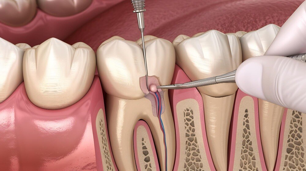
An infected tooth develops when bacteria pass through damaged enamel and dentine into the pulp, the living tissue at the centre. Deep decay, a cracked tooth, trauma, or repeated dental work or fillings can create that route.
Once the pulp becomes inflamed or dies, bacteria can travel through the root canals into the surrounding bone and gum.
These signs require a dental assessment:
| Symptom | What it can indicate | What to do |
|---|---|---|
| Lingering hot or cold sensitivity | Inflamed pulp that is not recovering | Arrange an examination |
| Spontaneous throbbing or pain when biting | Pulp inflammation or infection around the root | Seek prompt assessment |
| Gum swelling or a pimple on the gum | Drainage from an infected tooth | Book dental care promptly |
| Facial swelling or fever | Infection spreading beyond the tooth | Seek urgent dental care |
| A darkened tooth | Loss of pulp vitality or previous trauma | Arrange an examination |
Pain stopping does not prove that the tooth has healed. When the pulp dies, its nerves can no longer produce the original toothache, but bacteria and infection can remain inside the canals and continue damaging nearby tissues. A quiet tooth can therefore still need treatment.
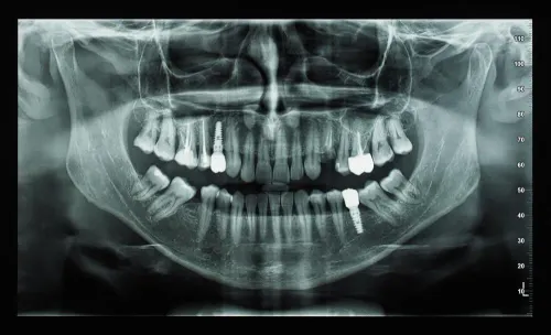
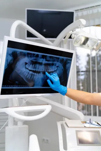
Increasing swelling, fever, difficulty swallowing or breathing, or severe pain that does not settle needs prompt assessment. A dentist confirms the cause with your history, examination, pulp-sensitivity and biting tests, and dental X-rays; a toothache alone does not prove that root canal treatment is required.
How the dentist confirms which tooth needs treatment
Root canal diagnosis begins with the pain history: which tooth hurts, whether heat or cold lingers, and whether biting triggers a sharp or dull response. The dentist then compares neighbouring teeth using a pulp-sensitivity test, taps each tooth with a percussion test, and uses a biting test for tooth pain.
No single result proves that root-canal treatment is necessary.
| Cause | Findings that point towards it | Useful imaging |
|---|---|---|
| Infected or dead pulp | Lingering thermal pain, spontaneous pain, tenderness to tapping, or no response to sensitivity testing | A targeted dental X-ray shows deep decay, widening around the root tip, or bone loss; early infection can still look normal |
| Cracked tooth | Brief, sharp pain when releasing a bite, with inconsistent cold sensitivity; a crack may not appear on an X-ray | An intraoral X-ray can exclude decay or bone changes, but magnification and crack-specific tests often matter more |
| Gum abscess | A tender, localised gum swelling or pus beside the tooth, with the tooth sometimes responding normally to sensitivity testing | An intraoral X-ray may show bone loss along the root surface rather than disease centred at the root tip |
| Neighbouring tooth or sinus pain | The suspected tooth tests normally, while another tooth reproduces the pain; sinus pain may affect several upper teeth with congestion or facial pressure | A Digital OPG surveys multiple teeth and the upper jaw; an intraoral X-ray gives finer detail for one tooth |
Sinus pain does not justify a root canal without matching dental findings. The dentist combines the examination, test results and images before choosing treatment; an X-ray alone cannot measure pulp vitality.
What happens during a painless root canal appointment
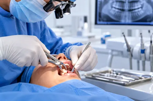
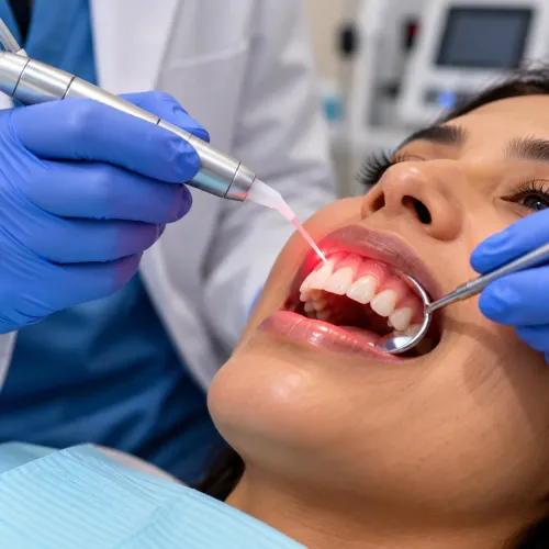
A painless root canal treatment starts with local anaesthetic around the tooth. The dentist tests the numbness before working; sharp pain is not something you must tolerate, so report it immediately and the dentist can pause and give more anaesthetic.
- The dentist reviews the examination and X-ray, then gives local anaesthetic.
- A rubber dam stretches around the tooth, keeping saliva out and protecting your mouth from instruments and cleaning fluids.
- The dentist makes an access opening in crown to reach the pulp chamber.
- Inflamed pulp removal clears the damaged or infected tissue. This removes pulp, not the tooth’s roots; your natural tooth stays in place.
- Cleaning and shaping root canals uses small files and disinfecting solutions to remove debris and create space for the filling.
- The dentist completes filling and sealing canals, then closes the access temporarily or permanently.
You should feel pressure, pushing, cool water, or vibration from the handpiece and canal instruments, but not sharp pain. The rubber dam can feel tight around the tooth, and keeping your mouth open may become tiring.
Treatment can take one appointment or several, depending on canal anatomy, infection and whether medication is placed inside the canals. Afterward, tenderness when biting can last for a few days; increasing swelling or severe pain requires prompt dental review. A final restoration, such as a filling, onlay or crown, protects the weakened tooth from fracture.
What improves treatment quality and when extra care is needed
Treatment quality depends on controlling the canal’s length, shape and bacterial load. Errors in any of these areas can leave infected tissue behind, push instruments beyond the root or create a seal that fails early.
- Electronic working-length measurement locates the canal endpoint more accurately than relying on an X-ray alone. This helps prevent over-cleaning near the tissues beyond the root.
- Magnification in root canal treatment helps the dentist identify small canals, cracks, calcifications and hidden openings that are easy to miss without enhanced vision.
- Rotary instruments use continuous motion to shape canals efficiently; reciprocating instruments use alternating motion and can reduce torsional stress in selected cases. The correct choice depends on canal curvature, width and instrument control.
- Canal disinfection uses irrigation and cleaning to reduce bacteria that instruments cannot reach. Shaping without thorough disinfection can leave infection in side spaces and microscopic irregularities.
One or more root canal appointments may be appropriate. Multiple visits become relevant when canals are complex, drainage is needed, infection is extensive or medication must remain inside the canals before sealing; this does not automatically mean treatment failed.
Antibiotics for dental infection do not remove infected pulp or replace drainage, root-canal treatment or extraction. They become relevant when infection causes systemic illness or spreads, such as fever or malaise. Seek urgent review for increasing facial swelling, fever, difficulty swallowing or breathing, or severe pain that does not settle.
How to protect the tooth after the canals are sealed
A sealed canal does not make the tooth immune to decay, gum disease or fracture. The access opening, old fillings, cracks and lost tooth structure can weaken it, so tooth restoration after root canal should be planned promptly rather than postponed until the tooth feels normal.
Posterior teeth often need cuspal coverage because chewing forces act on the remaining walls. An onlay covers selected cusps when enough sound tooth remains; a dental crown after RCT surrounds the tooth when it needs broader protection.
Your dentist should base the choice on remaining tooth structure, cracks, bite forces and tooth location—not on a standard package. Good tooth fracture prevention protects root-filled tooth function.
Continue brushing twice daily with fluoride toothpaste, use interdental cleaning, and attend examinations to detect new decay or gum problems. Post-treatment tenderness when biting can last a few days; increasing swelling, fever, trouble swallowing or breathing, or severe pain needs prompt review.
When comparing dental care in Paschim Vihar, ask these questions:
- Does the clinic assess remaining tooth structure before recommending an onlay or crown?
- Will you receive a written restoration plan, expected timing and review schedule?
- Can the same team check your bite, gums and X-rays if symptoms persist?
- What urgent support is available if swelling or severe pain develops?
Dr Batra's Dental is one clinic you can include in that comparison, using these practical questions rather than judging care by a painless-treatment claim alone.
| Care feature | What to compare | Why it matters |
|---|---|---|
| Restoration planning | Onlay versus crown recommendation | Matches protection to remaining tooth |
| Follow-up | Examination and X-ray review | Detects decay, gum disease or failure |
| Aftercare | Hygiene advice and urgent access | Protects healing and long-term function |
Related service
Root Canal Treatment (RCT) Restore your tooth health and protect your smile with painless root canal treatment at Dr Batra's Dental. View service → |
Frequently asked questions
How can a tooth remain infected after the pain disappears?
An infection can damage or kill the pulp, reducing pressure and nerve signals while bacteria remain inside the tooth and surrounding tissues.
How does a dentist confirm which tooth needs a root canal?
The dentist compares your symptoms with a clinical examination, pulp-response tests, tapping or biting tests, and dental X-rays.
Is root canal treatment painful?
Local anaesthetic numbs the tooth and surrounding area, so you should feel pressure rather than sharp pain during treatment.
How do you protect a tooth after root canal treatment?
Avoid chewing hard foods on the treated tooth until it is restored, maintain daily brushing and flossing, and complete the recommended permanent filling or crown.